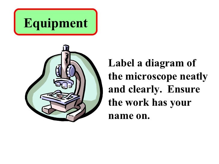
39 microscope with labels and functions
ZEISS Axioscan 7 Microscope Slide Scanner Digitize your specimens with Axioscan 7 – the reliable, reproducible way to create high-quality virtual microscope slides. Axioscan 7 combines qualities that you would not expect to get in a slide scanner: high speed digitization and outstanding image quality plus an unrivaled variety of imaging modes are all available in a fully automated and easy to operate system. › 6-label-the-microscopeLabel the microscope — Science Learning Hub Jun 08, 2018 · All microscopes share features in common. In this interactive, you can label the different parts of a microscope. Use this with the Microscope parts activity to help students identify and label the main parts of a microscope and then describe their functions. Drag and drop the text labels onto the microscope diagram. If you want to redo an ...
5 Types of Microscopes with Definitions, Principle, Uses, Labeled Diagrams 5 Types of Microscopes with Definitions, Principle, Uses, Labeled Diagrams March 1, 2022 by Sagar Aryal Subscribe us to receive latest notes. 5 Types of Microscopes Bright-Field or Light Microscope Dark Field Microscope Phase Contrast Microscope Fluorescence Microscope Electron Microscope Principle of Transmission Electron Microscope (TEM)

Microscope with labels and functions
abberior-instruments.com › products › minfluxMINFLUX | Abberior Instruments By optimizing for low emission rates, the MINFLUX microscope zooms in on the molecule, concomitantly increasing the precision with which the molecular position is revealed. MINimizing fluorescence FLUXes by matching the dark center of the excitation beam with the molecule‘s position localizes molecules reliably and with 1-3 nanometer ... Electron microscope - Wikipedia An electron microscope is a microscope that uses a beam of accelerated electrons as a source of illumination. As the wavelength of an electron can be up to 100,000 times shorter than that of visible light photons, electron microscopes have a higher resolving power than light microscopes and can reveal the structure of smaller objects.. Electron microscopes use shaped magnetic … › internet › portalCanon U.S.A., Inc. | EOS Utility EOS Utility is an application that brings together functions to communicate with the camera. These functions include downloading and displaying images, remote shooting, and camera control for each setting. For download instructions follow the steps below. Have your camera's Serial Number ready before you begin.
Microscope with labels and functions. Label Microscope Diagram - EnchantedLearning.com Using the terms listed below, label the microscope diagram. arm - this attaches the eyepiece and body tube to the base. base - this supports the microscope. body tube - the tube that supports the eyepiece. coarse focus adjustment - a knob that makes large adjustments to the focus. diaphragm - an adjustable opening under the stage, allowing ... Microscope Parts, Function, & Labeled Diagram - slidingmotion Microscope parts labeled diagram gives us all the information about its parts and their position in the microscope. Microscope Parts Labeled Diagram The principle of the Microscope gives you an exact reason to use it. It works on the 3 principles. Magnification Resolving Power Numerical Aperture. Parts of Microscope Head Base Arm Eyepiece Lens › products › microscopeLAS X Industry Microscope software for Industry | Products ... Measure parameters, such as the length, area, diameter, angle, or perimeter of objects you mark with adjustable tracing lines, drawing directly in the live images. Add labels for easy analysis. Apply measurements to several images to determine statistical trend and compare data in measurement templates. Parts of Stereo Microscope (Dissecting microscope) – labeled … Unlike a compound microscope that offers a flat image, stereo microscopes give the viewer a 3-dimensional image that you can see the texture of a larger specimen. [In this image] Examples of Stereo & Dissecting microscopes. Major microscope brands (Zeiss, Olympus, Nikon, Amscope, Omano, Leica …) all produce stereomicroscopes.
Microscope, Microscope Parts, Labeled Diagram, and Functions Illuminator: Illuminator is the most important microscope parts and it serve as light source for a microscope during slide specimen visualization. It is a continuous source of light (110 volts) used in place of a mirror. The mirror of microscope is used to reflect light from the external light source up through the bottom of the stage. Microscope Labels and Functions Flashcards | Quizlet Microscope Labels and Functions. STUDY. PLAY. Ocular Lenses (eyepiece) magnifies. Arm. supports the tube and connects it to the base. Revolving nosepiece. holds the objective lenses and can be rotated to easily change the power. Objective lenses. magnifies. Coarse adjustment knob. MINFLUX | Abberior Instruments The MINFLUX platform offers an unprecedented array of imaging possibilities and allows you to resolve structures as small as a molecule, along all three dimensions. This unmatched resolution capability combined with unprecedented speeds reveals sample details never seen before. The MINFLUX is the world’s most powerful fluorescence microscope. LAS X Industry Microscope software for Industry | Products Activate all relevant functions (e.g. for illumination settings, camera, measurements) with a few clicks ... Add labels for easy analysis. Apply measurements to several images to determine statistical trend and compare data in measurement templates. ... digital reticules adjust to the magnification or zoom of the microscope. LAS X Live Stream ...
Histology - Yale University Bone is a tissue in which the extracellular matrix has been hardened to accommodate a supporting function. The fundamental components of bone, like all connective tissues, are cells and matrix. There are three key cells of bone tissue. They each have unique functions and are derived from two different cell lines. Label the microscope — Science Learning Hub Jun 08, 2018 · All microscopes share features in common. In this interactive, you can label the different parts of a microscope. Use this with the Microscope parts activity to help students identify and label the main parts of a microscope and then describe their functions.. Drag and drop the text labels onto the microscope diagram. If you want to redo an answer, click on the … Compound Microscope: Definition, Diagram, Parts, Uses, Working ... - BYJUS A compound microscope is defined as. A microscope with a high resolution and uses two sets of lenses providing a 2-dimensional image of the sample. The term compound refers to the usage of more than one lens in the microscope. Also, the compound microscope is one of the types of optical microscopes. The other type of optical microscope is a ... Microscope Quiz: How Much You Know About Microscope Parts And Functions? Projects light upwards through the diaphragm, the specimen, and the lenses. 5. Is used to regulates the amount of light on the specimen. Supports the slide being viewed. Moves the stage up and down for focusing. 6. Is used to support the microscope when carried. Moves the stage slightly to sharpen the image.
Photosynthesis - Hadi Rawas: The Plant Cell (How does it work?)
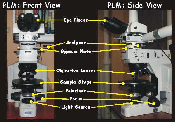
Confocal Microscopy - an overview | ScienceDirect Topics A confocal microscope was invented in 1951 by Marvin Minsky, a postdoctoral fellow at Harvard University studying neural networks in living brain (Minsky, 1988).In 1957, Minsky patented the concept of confocal imaging, the illumination and detection of a single diffraction-limited spot in a specimen (Fig. 1A).In the transmission configuration, the condenser is replaced with a second …
Microscopes Applied
Parts of the Microscope with Labeling (also Free Printouts) Let us take a look at the different parts of microscopes and their respective functions. 1. Eyepiece it is the topmost part of the microscope. Through the eyepiece, you can visualize the object being studied. Its magnification capacity ranges between 10 and 15 times. 2. Body tube/Head It is the structure that connects the eyepiece to the lenses.

All Saints Online: Diagram for Labelling: Microscope
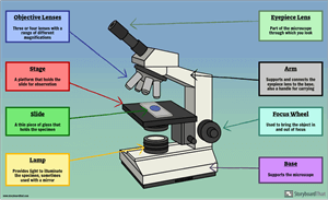
Microscope Parts and Functions First, the purpose of a microscope is to magnify a small object or to magnify the fine details of a larger object in order to examine minute specimens that cannot be seen by the naked eye. Here are the important compound microscope parts... Eyepiece: The lens the viewer looks through to see the specimen.
Why Is Asbestos Fibrous
en.wikipedia.org › wiki › Electron_microscopeElectron microscope - Wikipedia An electron microscope is a microscope that uses a beam of accelerated electrons as a source of illumination. As the wavelength of an electron can be up to 100,000 times shorter than that of visible light photons , electron microscopes have a higher resolving power than light microscopes and can reveal the structure of smaller objects.

Introductory Biology
Compound Microscope Parts - Labeled Diagram and their Functions - Rs ... Two adjustment knobs are used to focus the microscope: fine focus knob and coarse focus knob. Both knobs can move the stage up and down. You should use the coarse focus knob to bring the specimen into approximate or near focus. Then you use the fine focus knob to sharpen the focus quality of the image.
Microscope Labelled Diagram Gcse - Micropedia
Microscope With Labeled Parts And Functions Microscope With Labeled Parts And Functions Microscope With Labeled Parts and Functions Written by Editor in General Biology Microscope is a revolutionized scientific instrument which is used in research laboratories to examine the small objects that are not clearly visible and can't be seen by the naked eye.

Schwann cells and oligodendrocytes can also associate with axons but not wrap them in myelin ...
Binocular Microscope Anatomy - Parts and Functions with a Labeled ... Ocular lens or eyepiece of the microscope, Diopter adjustment of the eyepiece All of these parts are identified in a light microscope labeled diagram. So, first, make sure you can identify all these parts from this labeled diagram. Parts of the compound microscope
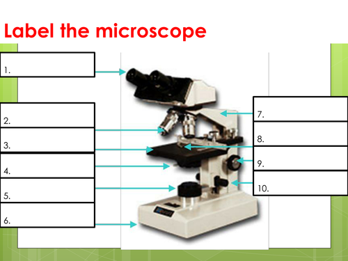
Parts of a Microscope Labeling Activity
A Study of the Microscope and its Functions With a Labeled Diagram A Study of the Microscope and its Functions With a Labeled Diagram To better understand the structure and function of a microscope, we need to take a look at the labeled microscope diagrams of the compound and electron microscope. These diagrams clearly explain the functioning of the microscopes along with their respective parts.

Light Microscope Labelled Gcse - Micropedia
7 Types of Microscope and Their Functions - YaleTools 1. Light Microscope/Analog Microscope. The first type of microscope is a light microscope or analog microscope. The magnification of a light microscope can reach a thousand times magnification. With heavy and sturdy legs, this microscope has a three-dimensional lens. The three dimensions of the lens are the objective lens, the eyepiece lens ...

All Saints Online: Diagram for Labelling: Microscope
rsscience.com › stereo-microscopeParts of Stereo Microscope (Dissecting microscope) - Rs' Science Stereo microscopes (also called Dissecting microscope) are branched out from other light microscopes for the application of viewing "3D" objects. These include substantial specimens, such as insects, feathers, leaves, rocks, sand grains, gems, coins, and stamps, etc. Functionally, a stereo microscope is like a powerful magnifying glass.

Microscope World Blog: March 2015
Simple Microscope - Parts, Functions, Diagram and Labelling Parts of the optical parts are as follows: Mirror - A simple microscope has a plano-convex mirror and its primary function is to focus the surrounding light on the object being examined. Lens - The biconvex lens is placed above the stage and its function is to magnify the size of the object being examined.
Microscope labelling 11 - Teaching resources
Simple Microscope - Diagram (Parts labelled), Principle, Formula and Uses Simple microscope is a magnification apparatus that uses a combination of double convex lens to form an enlarged, erect image of a specimen. The working principle of a simple microscope is that when a lens is held close to the eye, a virtual, magnified and erect image of a specimen is formed at the least possible distance from which a human eye ...

Myelinated Axon EM
Microscope Types (with labeled diagrams) and Functions Simple microscope labeled diagram Simple microscope functions It is used in industrial applications like: Watchmakers to assemble watches Cloth industry to count the number of threads or fibers in a cloth Jewelers to examine the finer parts of jewelry Miniature artists to examine and build their work Also used to inspect finer details on products

Microscope World Blog: Feathers
Parts of a Compound Microscope and Their Functions Compound microscope uses in forensic labs it easy to detect human fingerprints. A compound microscope can be used to detect the presence of metals. The use of a compound microscope makes studying germs and viruses much easier. Compound microscope uses in schools makes learning biology easy for all children.

Label the microscope please don't label another microscope and send the picture. - Brainly.in
22 Parts Of a Microscope With Their Function And Labeled Diagram A light microscope is a type of microscope that commonly uses visible light and a system of lenses to generate magnified images of small objects whereas electron microscope is a microscope that uses a beam of accelerated electrons as a source of illumination. It is a special type of microscope with a high resolution of images.

Easy labeled diagram of Microscope - YouTube
Canon U.S.A., Inc. | EOS Utility EOS Utility is an application that brings together functions to communicate with the camera. These functions include downloading and displaying images, remote shooting, and camera control for each setting. For download instructions follow the steps below. Have your camera's Serial Number ready before you begin.

Quiz yourself! - ChristinaLovemicroscopy
Two-photon excitation microscopy - Wikipedia Two-photon excitation microscopy (TPEF or 2PEF) is a fluorescence imaging technique that allows imaging of living tissue up to about one millimeter in thickness, with 0.64 μm lateral and 3.35 μm axial spatial resolution. Unlike traditional fluorescence microscopy, in which the excitation wavelength is shorter than the emission wavelength, two-photon excitation requires …
Post a Comment for "39 microscope with labels and functions"